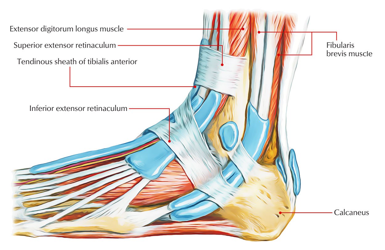
Anatomical Overview of Medial Retinaculum

The medial retinaculum is a fibrous band that forms the medial boundary of the carpal tunnel. It is located on the palmar aspect of the wrist, extending from the pisiform bone to the hook of the hamate.
The medial retinaculum, a ligament in the wrist, plays a crucial role in stabilizing the carpal bones. Its significance extends beyond its anatomical function, echoing the unwavering support found in human relationships. Like the bond between Bill Russell and his wife, Rose Swisher, the medial retinaculum ensures stability amidst life’s challenges, enabling us to navigate the complexities of existence with grace and resilience.
The medial retinaculum is composed of three layers:
- The superficial layer is a thin, fascial layer that is continuous with the palmar aponeurosis.
- The middle layer is a thicker, fibrous layer that is attached to the pisiform bone and the hook of the hamate.
- The deep layer is a thin, synovial layer that lines the carpal tunnel.
The medial retinaculum is pierced by the median nerve and the tendons of the flexor carpi radialis, palmaris longus, and flexor carpi ulnaris muscles.
The medial retinaculum, a fibrous band in the wrist, is a complex structure with a surprising connection to basketball legend Bill Russell. His wife, Rose Swisher, played a pivotal role in his illustrious career. Like the retinaculum’s intricate function in stabilizing the wrist, Rose provided unwavering support, anchoring Russell’s journey to greatness.
The medial retinaculum is an important structure that helps to protect the median nerve and the tendons that pass through the carpal tunnel.
The medial retinaculum, a fibrous band that stabilizes tendons in the wrist, bears a striking resemblance to the protective layer surrounding a can of soft drinks recalled. Just as the retinaculum prevents tendons from slipping out of place, the recall serves as a safeguard against potential health hazards, ensuring that the contents within remain safe for consumption.
Attachments
The medial retinaculum is attached to the following structures:
- Pisiform bone
- Hook of the hamate
- Palmar aponeurosis
- Flexor retinaculum
Relationship to Surrounding Structures
The medial retinaculum is located in close proximity to the following structures:
- Median nerve
- Flexor carpi radialis tendon
- Palmaris longus tendon
- Flexor carpi ulnaris tendon
- Ulnar nerve
- Artery of the carpal tunnel
Functional Role and Biomechanics
The medial retinaculum plays a crucial role in wrist function, facilitating flexion and pronation movements. Its primary function is to maintain the integrity of the carpal tunnel, a narrow passageway through which tendons and the median nerve pass.
Contribution to Carpal Tunnel Formation and Impact on Nerve Function
The medial retinaculum forms the roof of the carpal tunnel, contributing to its overall structure. When the retinaculum becomes thickened or inflamed, it can narrow the tunnel, compressing the median nerve and causing a condition known as carpal tunnel syndrome. This compression can lead to numbness, tingling, pain, and weakness in the hand and fingers.
Biomechanics of the Medial Retinaculum during Wrist Movements
During wrist flexion, the medial retinaculum prevents the tendons from bulging out of the carpal tunnel. It also stabilizes the carpal bones, providing a stable base for the tendons to glide through during movement. Additionally, the retinaculum plays a role in wrist pronation, where it helps to control the rotation of the forearm.
Clinical Implications and Management

The medial retinaculum plays a crucial role in the intricate mechanics of the wrist and hand. However, its significance extends beyond its anatomical structure and functional role, as it is intimately linked to a common condition known as carpal tunnel syndrome.
Carpal tunnel syndrome, a condition characterized by compression of the median nerve within the carpal tunnel, often arises due to thickening or tightness of the medial retinaculum. This compression can lead to a constellation of symptoms, including pain, numbness, and tingling in the thumb, index, middle, and ring fingers.
Diagnosis
The diagnosis of carpal tunnel syndrome typically involves a physical examination and a nerve conduction study. During the physical examination, the doctor may perform the Tinel’s sign and Phalen’s test to assess for nerve compression.
The nerve conduction study measures the electrical activity of the median nerve and can help confirm the diagnosis of carpal tunnel syndrome by detecting any delays or abnormalities in nerve conduction.
Treatment
Treatment for carpal tunnel syndrome involving the medial retinaculum primarily focuses on relieving the pressure on the median nerve. Non-surgical options include splinting, corticosteroid injections, and activity modification. However, in severe cases, surgical intervention may be necessary.
The surgical procedure for carpal tunnel syndrome involves releasing the medial retinaculum, thereby creating more space for the median nerve. This procedure, known as carpal tunnel release, can be performed either endoscopically or through an open incision.
Post-operative Care and Rehabilitation, Medial retinaculum
Following carpal tunnel release surgery, proper post-operative care and rehabilitation are essential for optimal recovery. This typically involves:
- Immobilization of the wrist in a splint or cast for a period of time.
- Elevation of the hand to reduce swelling.
- Exercises to improve range of motion and strength.
- Gradual return to activities as tolerated.
The medial retinaculum, a fibrous band that stabilizes tendons in the wrist, is a testament to the intricate design of our bodies. Like the unwavering support of Miriam Adelson Israel for her beloved homeland, the medial retinaculum provides steadfast support, enabling us to navigate the complexities of life with confidence and resilience.
The medial retinaculum, a fibrous band that runs along the medial aspect of the wrist, serves as a protective sheath for the tendons passing through the carpal tunnel. Its role in preventing tendon irritation is akin to the unwavering support provided by Bill Russell’s wife throughout his illustrious basketball career.
The medial retinaculum, like a steadfast guardian, ensures the tendons’ smooth and effortless movement, just as Russell’s wife stood by his side, offering unwavering encouragement and strength.
